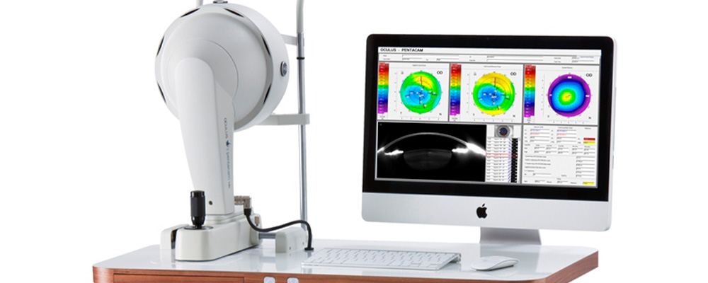
What is Corneal Topography?
Corneal topography is an imaging technique that maps the surface of the cornea, the transparent front layer of the eye. This test provides detailed information about the shape, curvature and irregularities of the cornea.
Because the cornea is an important optical component of the eye, corneal topography can assist ophthalmologists in diagnosing and treating a variety of eye problems.
In Which Situations Can Corneal Topography Be Applied?
Evaluation Before Laser Refractive Surgery
Corneal topography is performed especially before laser refractive surgical procedures such as LASIK. This can help identify suitable candidates for surgery and help plan surgery.
Astigmatism Evaluation
Astigmatism is a condition in which the cornea has an irregular shape. Corneal topography can be used to determine the amount and location of astigmatism.
Keratoconus Follow-up
Keratoconus is a condition in which the cornea takes a conical shape. Corneal topography is important for early diagnosis and follow-up of keratoconus.
Contact Lens Compatibility
Corneal topography can help select the appropriate contact lens by evaluating a person’s eye structure.
Corneal Diseases and Injuries
In cases of corneal injuries or diseases, corneal topography can help doctors understand these conditions.
Corneal topography is generally a simple, quick and painless test. Patients usually sit in front of the device while their eyes look at a fixed point.
The topographic map shows the surface of the cornea in color, which allows the doctor to evaluate the irregularities and shape of the cornea. This information can help the ophthalmologist create a treatment plan or prescribe appropriate glasses or contact lenses.
Create an Appointment Online
